 |
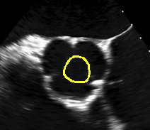
Aortic valve segmentation based on GVF-snake
The aortic valve is significant in image-guided cardiac intervention and therapy. The GVF-snake is used for the segmentation of aortic
valve in US image for the following reasons: 1) the cardiac valve has several boundary concavities and GVF-snake can converge to these
concavities. 2) GVF-snake allows a simple initialization of the snake since it has a large capture range, which can reduce the user
interaction. In this project, user need only draw a small circle in the center of the image as the initial counter and it will converge
after less than 20 iterations. The initial circle can also be automatically generated. As a result, accurate segmentation can be obtained
without user interactions. (C++ implementation)
|
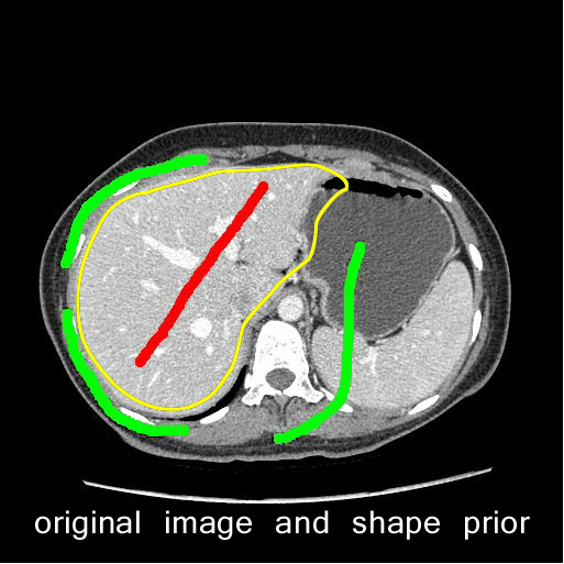 |
Liver segmentation using Graphcut with shape prior
Graphcut has been widely used in medical image segmentation. However, since adjacent organs have similar grayscales to those of liver,
many segmentation methods can easily lead to over-segmentation. In this project, I tried to incorporate a shape prior into the
graph cut method. The shape prior is employed to construct the weight of each edge, so that a shape energy is defined and incorporated in
the energy function. Compared with the initial graphcut without shape prior, this method can obtain a smoother counter and exclude
adjacent organs. Max-flow is used to solve the optimization problem.(C++ implementation)
|
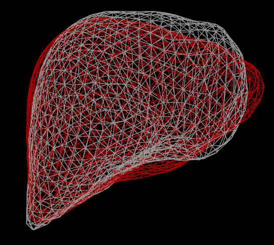 |
Shape correspondence in statistical shape model
Point correspondence between shapes play a critical role in ensuring the performance of statistical shape model.
I tried different methods, including ICP, TPS-RPM, SPHARM, and AFDM. In my application of modeling liver shapes,
which have a complex variation, I found AFDM has a better performance.(MATLAB implementation)
|
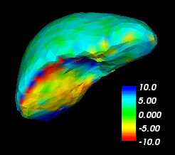 |
Toolkit for image segmentation evaluation
I developed a toolkit to facilitate the evaluation of medical image segmentation. Evaluation based on metrics including Dice coefficient,
average symmetric surface distance (ASD), and Hausdorff distance were implemented. All the quantitative evaluation could be achieved by
this toolkit. In addition, distance errors were visualized to display the mis-segmentation. For example, in the left figure, the shortest
Euclidean surface distance was measured in millimeters and visualized. Blue color shows over-segmentation and red color shows under-segmentation.
|
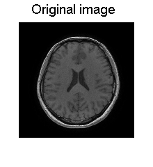 |
Image reconstruction using compressed sensing
Medical images exhibit transform sparsity, and they have a sparse representation in terms of spatial finite differences,
their wavelet coefficients, or other transforms. In this project, a non-uniform random sampling method was used to
measuring only a subset of image’s k-space. The wavelet coefficients, which could be treated as a sparse signal,
were reconstructed from the unsampled image’s wavelet transform using L1 penalty, solved by iterative soft-thresholding.
Then the initial image can be reconstructed by the wavelet coefficients. (MATLAB implementation)
|
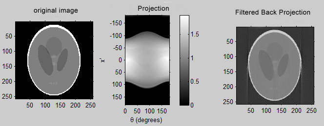 |
Radon transformation and Filtered Back Projection
I write a function to generate the Shepp-Logan phantom, the user can adjust different parameters such as Xoff, Yoff, Dx, Dy, Nx and Ny,
so that images in different sizes and resolutions can be obtained. Then I write a function to realize Radom transformation. In addition,
the projection data can also be acquired by using Joseph algorithm and blob approach, which can be compared with the result of Radom
transformation. To backproject the data, a ramp filter is implemented on the projections and then the results are back projected.
(MATLAB implementation)
|
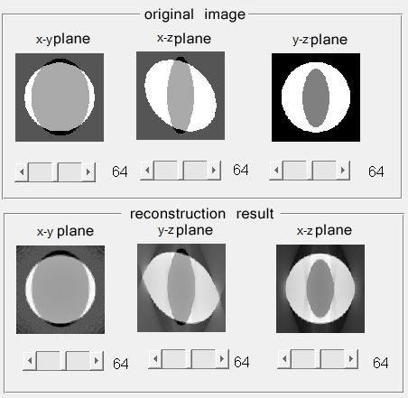 |
Simulations on cone-beam CT reconstruction
In this project, I simulated the 3D cone-beam CT reconstruction. The whole progress consists of four parts:(MATLAB implementation)
- generate 3D phantoms with arbitrary ellipsoids
- generate cone-beam data
- filter the data with H_ramp(t)
- back project the filtered projection data
|
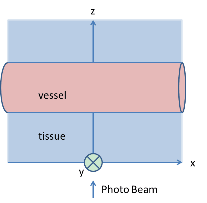 |
Monte Carlo modeling of light transport in tissue with vessels
Monte Carlo simulations offer a flexible approach to photon transport in turbid tissues. It is based on macroscopic optical properties
of small units of tissue volume. The most famous related work is that of
Lihong Wang,
who modeled the photon transport in multi-layered
tissues. In this project, the Monte Carlo simulation deals with the transport of an infinitely narrow photon beam, perpendicularly incident
on a tissue under which a vessel is parallel to the surface of the tissue. The tissue, which is infinitely wide and deep, and the vessel,
which is infinitely long are described by the refractive index, the absorption coefficient, the scattering coefficient, and the anisotropy factor.
This simulation can help a better understanding of the optical imaging of tissue with vessels. This work was mentored by
Shanbao Tong.
|
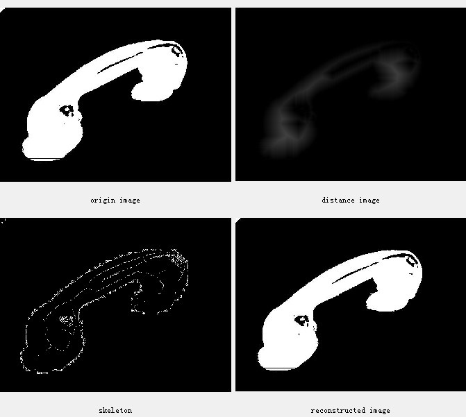 |
Digital image processing toolkit
In this project, I implemented an image processing software which can handle a lot of tasks: (C++ implementation)
- histogram analysis and segmentation based on threshold value (Otsu, Entropy)
- morphological operation such as opening and closing
- filtering and noise deducting
- edge detection (use different kernels such as sobel, pewitte, Roberts and user defined kernel)
- distance transform and skeletonization, followed by reconstruction
|
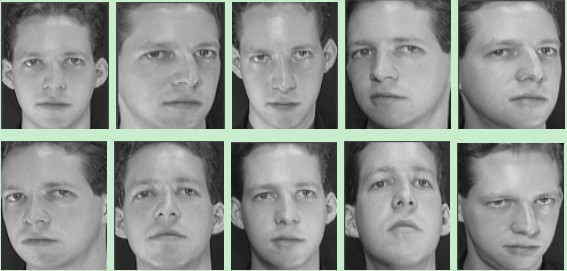 |
Face recognition
This project is the dissertation for my second major (Intelligence Science and Technology) when I was an undergraduate student.
This project studied the application of texture features in face recognition and texture features based on gray level co-occurrence
matrix (GLCM) from different directions were used. The correlation extracted from GLCM based on different offset in the same
direction was fitted in to cubic polynomial curve and the factors of the curve were used to construct feature vectors. In the last step,
classifiers based on support vector machine (SVM) were employed for identification. This work was mentored by
Jingqi Yan.
|