 |
Liver segmentation
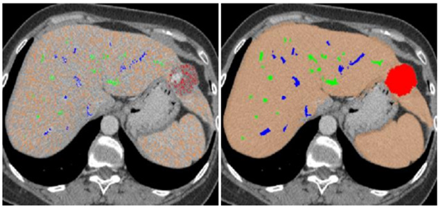
We proposed a minimally supervised classification method to segment hepatic parenchyma (with or without tumors), portal veins,
and hepatic veins from contrast enhanced CT abdominal data for living donor liver transplantation surgery planning and
guiding system. This method employs both statistical and spatial information to overcome the ambiguities caused by
the overlap in intensities among different tissue classes as well as the ambiguities due to noise and partial volume effect.
|
 |
Liver shape modeling
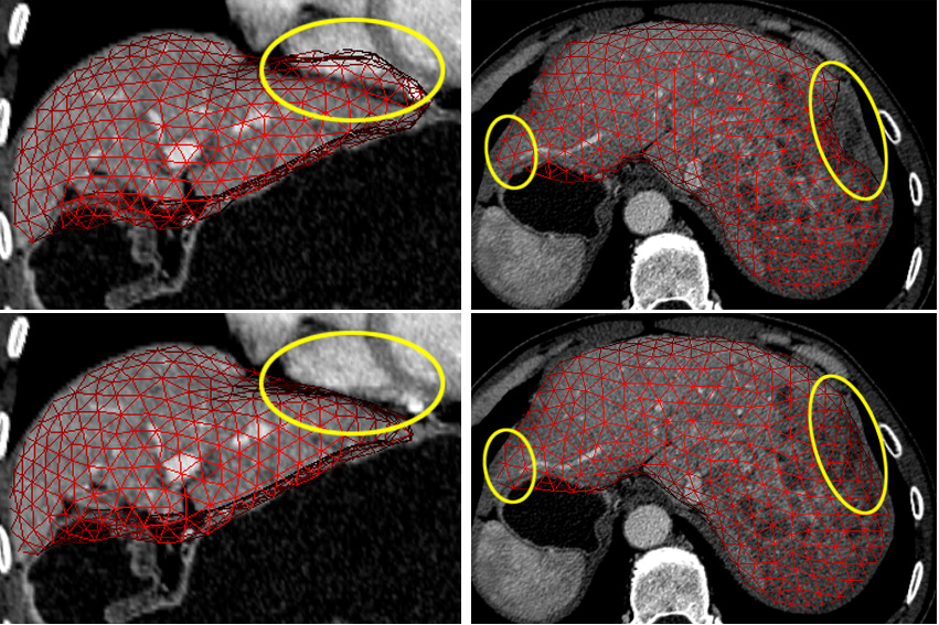
Many Statistic Shape Modeling (SSM) methods have been widely used to model the shape variation, among which the active
shape model (ASM) and its variations are the most prevalent. However, the modeling of liver shapes of patients with liver
cancer is much more challenging than that of many other organs. We use the sparse shape composition (SSC) to implicitly m
odel the liver shape. SSC represents the shape by a sparse linear combination of shapes in the shape repository. It does not
rely on any assumption of parametric distribution models. SSC can also preserve the local detail information that presents
in the training set.
|
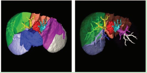 |
Liver surgery planning system
To achieve the best resection plan for liver surgery, surgeons need to identify the location of the liver portion that would be cut off,
together with the distribution of intrahepatic vessels and tumors. As a result, the preoperative planning based on medical image is
highly important. Our liver surgery planning system consists of three basic modules:
- Segmentation of hepatic parenchyma, portal veins, hepatic veins and tumors.
- Liver segment approximation that divides the liver into several functionally independent segments.
- Visualization of segmentation result and liver segments.
We proposed a novel segmentation framework for this system. The new
framework use SSC to model the shape prior for the liver, then the shape prior is cooperated with the Fast Marching method to get
accurate segmentation. It not only obtains accurate segmentation results for healthy persons and common patients but can also
deal with patients with liver cancers in complex clinical environments.
|
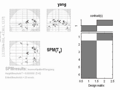 |
Evaluation of traumatic brain injury based on DWI and Voxel Based Analysis (VBA)
The microstructure of white matter may change when people suffer from traumatic brain injury, but this can hardly be detected by usual CT
and MRI. Taking advantage of TWI, we can find the abnormalities of the diffusion anisotropy and then assess the degree of injury.
Using the method of voxel based analysis (VBA) and Statistical Parametric Mapping (SPM), I built a two-sample t-test model after some
spatial pre-procession, in order to find regions where the fractional anisotropy (FA value) was obviously lower than that of normal person.
Finally abnormalities of white matter fibers can be located and the volume of injury can be calculated.
|